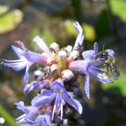
Dufourea bee on flower

pcyu_logo

By Sheila Dumesh, entomology research assistant at York University.

——————————–

My interest in bees was ignited in 2007, when I took a biodiversity course in my last year as an undergraduate student at York University in Toronto.  The course instructor was the well-known melittologist, Laurence Packer, and, although I had not met him before, I had heard many good things.  Laurence’s affection for bees was inspiring, not only to me, but to others in the past and many more to come.  He was so fascinated by these cute and fuzzy insects (at the time, I did not see myself describing them as such).  Even though he had been studying bees for decades, the look of excitement on his face never faded when collected and examined them.  Back then, my knowledge of bees was very limited.  I was unaware of their diversity, importance, and great beauty!

I began with an Honours thesis under Laurence’s supervision in the “bee lab” at York University.  I was keen on taxonomy and began a systematic study on a Central American bee genus, Mexalictus.  For my Master’s thesis, I chose to continue that work and complete a revision of Mexalictus, which included descriptions for 20 new species, an illustrated key, and a phylogenetic analysis.  I conducted my field work in Costa Rica, Guatemala, and Mexico, where I sampled in high elevation cloud forests (the known habitat of Mexalictus).  As these species are quite rare, I did not always have the pleasure of finding them; although this was somewhat upsetting, I was amazed by the bee (and general insect) diversity in that part of the world.  I was aware of it, but being out in the field in those countries was a truly amazing experience.  Just the change in habitat and species make-up along a small sector of the elevation gradient was incredible to witness!

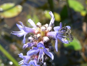
Dufourea bee on flower

Dufourea sp. – Photo by Sheila Dumesh

Throughout my time as a Master’s student, I studied other groups of bees and collaborated with others in our lab.  One such project is the revision of the Canadian species in the genus Dufourea (Apoidea: Halictidae), which I undertook with Cory Sheffield and recently published in the Canadian Journal of Arthropod Identification.  There are eight species in Canada, but some were described from only one sex, the descriptions were written by several authors in different publications, and a key to identify these species was previously unavailable!  These bees are also floral specialists, meaning they visit specific flowers (usually a genus or family).  Cory and I set out to revise this group and provide all of this information in one paper.  The identification key is user-friendly and illustrates the characters mentioned in the key couplets to aid the user.  We also constructed species pages, which include full descriptions, important features, distribution maps, and images of each species.

We are striving towards creating many more illustrated (and web-based) keys to facilitate bee identification.  I am very excited to have this work freely available and hope that it is found useful by others in the community!

———–

Dumesh, S. & Sheffield, C.S. (2012). Bees of the Genus Dufourea Lepeletier (Hymenoptera: Halictidae: Rophitinae) of Canada, Canadian Journal of Arthropod Identification, 20 DOI: 10.3752/cjai.2012.20

Honey bee flying with pollen - Photo by Alex Wild

Honey bee flying with pollen – Photo by Alex Wild, used with permission

Honeybee colonies are famous for their orderly divisions of labour.  As worker bees grow up, they transition from housekeepers (cleaning the colony) to nurse bees (feeding young bees), to finally switching to foragers who go out and collect nectar and pollen for the rest of the colony.   To maintain a healthy colony, bees need to decide how many foragers and how many nurse bees are needed, and control of these numbers is accomplished by pheromone levels within the colony.

In honeybee colonies, there are pheromones like the alarm pheromone that cause immediate behavioural responses (called releaser pheromones) and others that trigger physiological changes like hormones do (called primer pheromone).  From previous work, it seemed that ethyl oleate functions as a primer pheromone, produced by foragers, that delays the maturation of nurse bees into foragers.

“Ethyl oleate does not elicit any noticeable behavourial responses in recipient workers,” says Dr. Erika Plettner, who supervised a recent study on the synthesis of ethyl oleate at Simon Fraser University in British Columbia.  “Yet it has a profound physiological effect”.

To understand how this chemical is produced in the individual bee and then distributed in the colony, Carlos Castillo and colleagues from Simon Fraser University in British Columbia and the Laboratoire Biologie et protection de L’Abeillie in France looked at several ways to identify the source and synthesis of ethyl oleate.  This chemical can be produced by a reaction between oleic acid (a common fatty acid in insects) and ethanol.  While you might not think of honeybees as heavy drinkers, it turns out that yeasts in flower nectar ferment the sugars present into ethanol, and so the forager bees have much higher exposure to ethanol than nurse bees.

To figure out if ethanol and oleic acid can be made into ethyl oleate by honeybees, the researchers incubated different honeybee body parts from forager and nurse bees with these precursors.  They found highest production of ethyl oleate in the head tissues, and that both nurses and foragers could produce ethyl oleate when given ethanol.  In addition, in whole bees, they found that the ethyl oleate migrated from the gut to the exoskeleton of the bees where it would exude into the colony.

Taken together, these results suggest that making ethyl oleate, while it is useful for colony control, might also be a way to deal with the occupational hazard of consuming toxic ethanol.  “Foragers have much higher occupational exposure to ethanol than nurses do,” says Dr. Plettner.  “This is why they make ethyl oleate in nature”.

Ethyl oleate molecule

Ethyl oleate

To track down where exactly the ethyl oleate was synthesized, they coupled oleic acid to a chemical that would produce fluorescence when the oleic acid was combined with ethanol to produce ethyl oleate.  Under the microscope, areas that fluoresced showed where ethyl oleate was being made.  They found that ethyl oleate was made in the esophagus, honey crop and stomach.

The authors were also able to identify the genes responsible for the synthesis of ethyl oleate in the honeybee and the resulting enzymes that catalyze the reaction between oleic acid and ethanol.  These enzymes are then secreted into the gut fluid, where they produce ethyl oleate, which is then transported to the cuticle.

The biosynthesis of ethyl oleate then can be thought of a way of providing updates to the colony about the availability of flower nectar in nature.  “EO might be some kind of ‘resource meter’ that tells the nurses in the colony how many nectar and pollen resources are coming in,” says Dr. Plettner.  “If lots of food is coming in, then it makes sense to inhibit nurse to forager transition, as the nurses would be more needed in the brood chamber than as foragers.  Conversely, if few resources and/or foragers are coming in, then it makes sense to speed up development of nurses so that they can forage and fill the need.”

Castillo, C., Chen, H., Graves, C., Maisonnasse, A., Le Conte, Y. & Plettner, E. (2012). Biosynthesis of ethyl oleate, a primer pheromone, in the honey bee (Apis mellifera L.), Insect Biochemistry and Molecular Biology, 42 (6) 416. DOI: 10.1016/j.ibmb.2012.02.002

Corresponding author: Erika Plettner (plettner@sfu.ca)

Further reading:

Castillo, C., Maisonnasse, A., Conte, Y.L. & Plettner, E. (2012). Seasonal variation in the titers and biosynthesis of the primer pheromone ethyl oleate in honey bees, Journal of Insect Physiology, 58 (8) 1121. DOI: 10.1016/j.jinsphys.2012.05.010

Pollenia griseotomentosa Calliphoridae Cluster fly
Pollenia rudis Face

Pollenia rudis

By Adam Jewiss-Gaines,  a research assistant at Brock University.

———————

When people ask me what the heck a calliphorid is (often after I have mentioned the family name and am being gawked at as if I’m crazy), I usually remark “You know those shiny flies you often see flying around in the spring and summer?”  This isn’t technically 100% accurate since the genus Pollenia, one of the most commonly encountered genera of the family, is in fact non-reflective and grey.  Upon closer inspection, a keen eye can also observe varying amounts of wrinkled, yellow hairs on the thorax.  These two qualities distinguish Pollenia from other blow flies throughout North America.  Despite being a little dull when compared to their more eye-catching iridescent relatives, Pollenia are ecologically important insects as they aid in plant pollination and the processing of various biomaterials.

Pollenia often become particularly active during the spring and summer months once the temperature warms up, although they can occasionally be spotted indoors in the wintertime on a warmer day.  With a sudden onslaught of large, grey insects flying around when the snow begins to melt, it comes as no surprise that people tend to get irritated with them and consider them pests.  Oftentimes they are mistaken as houseflies (Family Muscidae) causing Pollenia species to be labeled as potential food contaminators, but this is not the case.  These insects are also particularly well-known for their clustering behaviour on walls, earning them their common name: cluster flies.

Even though Pollenia are extremely common, their general biology is largely unknown with a few exceptional details. It is known that larval Pollenia are parasites on various other organisms, such as maggots and worms. For example, Rognes (1991) noted that Pollenia pediculata, one of the most common species found throughout the continent, is a parasite of the earthworm species Eisenia rosea. Aside from this little tidbit however, specific information regarding the life cycles of Pollenia species is relatively scarce and further studies in this particular field would greatly improve our knowledge of the genus.

Pollenia griseotomentosa Calliphoridae Cluster fly

Pollenia griseotomentosa

Until very recently it has been thought that all Pollenia found in North America were the same species (Pollenia rudis), but after examining various collections throughout the world, Knut Rognes found that six members of the genus occur throughout the region.  Terry Whitworth adapted much of Rognes’ work shortly thereafter into a nice, clean, simple identification key for North America. With accurate images and photography, however, characters could be even easier to distinguish and observe when one is able to compare a photograph to the creature they have under their microscope.

Therefore, to further expand on Terry’s key and clarify important visual characters, I collaborated with him and Dr. Steve Marshall to create a fully-illustrated digital key for distinguishing the six North American Pollenia species from one another.  Now published in the Canadian Journal of Arthropod Identification, Cluster Flies of North America couples high-resolution images of important traits with a clean and simple interface to create a handy tool to be used by entomologists and non-entomologists alike. If you are relying on this key for identification, it is recommended to use physical specimens of Pollenia rather than images or photos, since even the best of hand-photographs have difficulty capturing key features. In addition, distribution maps are provided for each species, constructed from locality data of specimens from the University of Guelph Insect Collection and Terry Whitworth’s personal collection of Pollenia.

Creating this key has been a great opportunity, and I hope the entomological community is able to make good use of it. My sincere thanks go out to Steve Marshall, Terry Whitworth, the editors, and my labmates and friends for all of their support.

——————————

Jewiss-Gaines, A., Marshall, S.A. & Whitworth, T.L. (2012). Cluster flies (Calliphoridae: Polleniinae: Pollenia) of North America, Canadian Journal of Arthropod Identification, 19 DOI: 10.3752/cjai.2012.19

Rognes, K. 1991. Blowflies (Diptera, Calliphoridae) of Fennoscandia and Denmark. Fauna Entomologica Scandinavica Vol. 24.

ESC Caption C1 P2

We had a great response to last week’s photo, so thank you to everyone who played along. We’ve got an all new photo for you to caption today, but first we need you to vote for your favourite Photo 1 caption.

ESC Caption Contest C1 P1

[polldaddy poll=6409189]

We’ll post the results and award some points next week.

Now, onto this week’s photo (here are the rules if this is your first time):

ESC Caption C1 P2

ESC Caption Contest C1 P2 – Photo by Morgan Jackson

Have fun!

Got a great insect photo? Submit it to the 3rd Annual BugEye Photo Contest presented by the Entomological Society of Ontario!

Acorn Weevil by Crystal Ernst

2011 Winning Photo, Open Category: Acorn Weevil by Crystal Ernst

Prizes for:
– Best photo (open category): $50
– Best photo by an Ontario resident: $50
– Best photo of an Ontario insect: $50
– Best photo by a kid under 13: $50

Open to everyone, no entry fee!
(Ontario resident includes anyone who currently makes their primary residence in Ontario, international students welcome!).

Submission deadline: Sept. 6th, 2012

Submit photos to: esophotos@gmail.com

Winners announced: September 30th, 2012

Copyright for the photo remains with the photographer, use must be granted for ESO promotional material. Winning photos will be displayed on the ESO website, and all entries will be displayed at the 149th Annual General Meeting of the ESO.

Interested in meeting other entomologists and learning more about Ontario insects? Join ESO! It’s free for students and amateurs, and only $30 for others. Get more information at http://www.entsocont.ca.

Rules:
1. Photos must be of insects or closely-related arthropods (e.g. mites, spiders).
2. Submissions must be as digital files
3. Photographic enhancement is allowed as long as it is something that could be achieved in a real darkroom (i.e. adjustment of contrast, color enhancement, cropping, etc.). However very obvious enhancements will be negatively scored.
4. You may submit up to 3 unique images per category.
5. Submit photos as 7.5 x 10 inches in size at 300 dpi (2250 x 3000 pixels), in .jpg format, with filename as title_lastname_firstinitial.jpg (e.g. dragonfly_smith_j.jpg).
6. Photos may be landscape or portrait in orientation.
7. Print photos must be scanned and submitted as digital files.

Please include a short description of your photo:
1. Where they were taken
2. Why you like them
3. What insect is pictured
4. What category is being entered
5. Your complete address

Judging criteria:
1. Image composition
2. Visual impact
3. Subject interest
4. Sharpness of subject
5. Difficulty of image acquisition
6. Depth of field within image

Dear Buggy is the the alter-ego of Dr. Chris MacQuarrie, a research entomologist with the Canadian Forest Service. You can ask Buggy questions of your own on Twitter @CMacQuar.

—————————-

Hello!

Dear Buggy has lept out of the pages of the ESC Bulletin and landed in the new and exciting wilderness of the ESC blog. My loyal readers shouldn’t worry, I’ll still be writing my column, but between editions of the Bulletin I’ll be posting here.

I’m very excited to be contributing to the ESC blog, but I’ll admit I am a tad nervous. When Crystal and Morgan invited me to contribute I was worried that it would be hard to come up with interesting topics. Thankfully the ideas began to flow after a glass of good scotch and I think I’ve come up with a few ideas that should keep me busy. After that? Well I’m always open to suggestions.

While thinking about this first blog post for ‘Dear Buggy’ I recalled how I felt when I first signed on to write Dear Buggy for the Bulletin. Where was I going to get these ideas!? Fortunately, a lot of the suggestions for my early columns come from the then-editor, Kevin Floate. Kevin had the original idea for Dear Buggy and shared with me his collection of questions and ideas. Later on, the ideas began to flow and inspiration came from others around me. Although, when I’m stuck for an topic I still go back to the original list that Kevin gave me. Good ideas can be hard to come by when you’ve got writer’s block and a deadline is fast approaching

As I planned this blog post I began to muse over the source of all my ideas, in particular “Where do I get my research ideas?”.

For example, when I was a new MSc student many of my research questions were influenced by the ideas of my supervisors. This isn’t all that unusual.  I suspect that when most of us started in research we were given, or at the least influenced, by ideas of others. As we mature scientifically we eventually start to come up with our own ideas. In fact, a good part of becoming a successful, independent researcher is tied to coming up with good ideas (which we might also call hypotheses). So where do these ideas come from? And perhaps more importantly, what do we do with these ideas once we have them?

I find inspiration hits at the oddest times and in the oddest places . I think Jorge Cham at PhD comics captured it best in this series of comics. Like most, I’ve been inspired in the ‘usual’ places: reading papers, attending seminars, talking with colleagues, etc… But inspiration can happen in other places as well. My mind tends to wander on my bike-ride home, when I’m pushing my daughter in her stroller, and quite often when I’m sharing a glass of scotch with my wife (who, lucky for me, is also an entomologist). As it turns out, this ‘mind wandering’ actually helps you have those ‘eureka’ moments, especially if you have been banging your head against the wall for awhile. I wrote recently about figuring out when you are best at writing. I think that advice can be extended to figuring out when and where you are inspired and to make sure you go there often.

But what about capturing ideas? My mind is like the proverbial sieve, but with one annoying quirk. I often can remember that I had an idea, I just can’t remember what it was.

To combat this selective memory I try to capture my ideas in my work journal as soon as possible. I’m a bit old fashioned so my journal is still kept in a notebook. Since my journal is also where I keep track my current projects, I make sure I highlight any new ideas so they are easy to find later on. There are many, many web-tools out there that can do the same job. The trick, though is to find something that works for you and to use it. My wife, for example,  is also an artist and long-ago got in the habit of carrying a sketchbook with her. That sketchbook now contains just as many ideas for research projects as it does ideas for art projects.

Finally, I must make a confession. Most of my ideas are bad. Some are half baked, others were thought of by someone else and rejected 30 years ago, a lot are impractical, infeasible, or near-impossible to execute or fit into the research that I’m doing. These over time get filtered out. Those that survive this process of natural selection, I keep. I then draw from this storehouse when the right moment comes along. Not all of these ideas will pan out of course, but by hanging on to the good ones I always have the right idea at hand when opportunity presents itself.

I’d be curious to hear about where you find your inspiration and how you track your ideas. Leave them in the comments section and I’ll summarize the best ones in a later post.

Cheers and see you next month,

Buggy.

By Crystal Ernst, PhD Candidate (McGill University)

Since I finally submitted my manuscript to a journal (YAY!), I’ve been tying up the little loose ends remaining at the end of the project. You know: organizing the useful data and image files, tossing the files marked “MESSING_AROUND_WITH_DATA_v.29), tidying up my R code, and, perhaps most importantly, curating my specimens.

I’m not going to go into too much detail about the project here (I’m saving that for another post). I will say, though, that the work I just completed includes just over 2,600 beetles from a single location in Nunavut (Kugluktuk, where I spent my entire first field season).

Two major aspects of the physical work (as opposed to the thinking, reading and writing) involved in an ecological/entomological project such as this one are the pinning and the identifications. Some of the tasks are a bit tedious (cutting labels; entering data; gluing over 800 specimens of the same tiny, plain black ground beetle to paper points), and some of them are thrilling (finally getting over the “hump” of the morphological learning curve and feeling good and confident when working with your keys; having experts tell you “Yep, you got those all right”; discovering rare species or new regional species records). In the end, in addition to the published (*knocks on wood*) paper, you have boxes or drawers full of specimens.

The specimens are gold. (Read this post by Dr. Terry Wheeler to understand why.)

Unfortunately, they don’t always get treated as such.

In the two-ish years that I’ve been working in my lab, we’ve had two major “lab clean-up days”. The first managed to get rid of a lot of clutter (old papers, broken apparatus, random crap). The second involved going through the “stuff” that was eating up all the most valuable storage space: specimens. Years and years worth of graduate and undergraduate projects’ specimens, stashed in freezers, boxes, bags and vials of all shapes and sizes.

Some things were in good shape (pinned well, or in clear ethanol). Other things were, well, downright nasty: gooey beetles in sludgy brown ethanol, dried up bits of moth wings in plastic containers, and a little bit of “what in the name of pearl is growing on that agar plate???” in the fridge.

None of these items were kept – their value as useful specimens was nil. So, the physical representation of some student’s work – probably months or years worth of work – was tossed in the trash.

Others, happily, were tucked back into drawers and cupboards, because someone had taken the time to ensure the specimens were well-preserved.

However, even many of these were suffering from a serious issue: bad labels.

Allow me to illustrate the point. This is a bad label:

This is also a bad label:
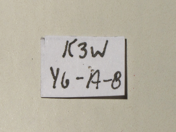
The first, you’ll note, is written in ballpoint pen (which fades) on a torn piece of notebook paper and contains almost no information. The second, although it looks fancier and perhaps more sciencey, is just as bad: it contains a cryptic code that is useful only to the bearer of the lab notebook in which said code has been written down. Or, perhaps the code is completely intelligible to the researcher who developed it, but the key to it exists only in his or her head.

To everyone else, it is meaningless. Neither of these labels indicate who collected the specimen, where, when, or how. And we all know what happens in labs: upon completion of their degrees, students move on, email addresses change, notebooks are misplaced, data files are not backed up. The labels’ codes can never be broken, and the scientific value of the specimens – *poof*.

While there’s nothing wrong, in theory, with using labels like these temporarily (although there is always a risk that they will be misinterpreted or misunderstood after a little while, even by the person who wrote them), they are absolutely useless as permanent records.

These are good labels:

These labels, properly affixed to a specimen, provide clear and universally understood information. One provides the location, including GPS coordinates, a method of collection, a date, the name of the collector(s). The information that goes on this label can vary a bit (it may include information about the habitat or host plant, for example), but those are the basic requirements. The smaller label is typically affixed on the pin below the first, and contains the specimen’s scientific name and the name of the person who identified it (it is the “det. label”, i.e., “determined by”). These labels, and therefore the specimen with which they are associated, will remain useful for decades, even centuries.

I am totally guilty of both of the offenses I just explained (the gooky vials of nastiness and the bad labels). For my undergraduate honors project, I identified close to 8000 spiders, mites and insects to the Family level – it was hundreds of hours of microscope work. Then I stuffed all those specimens back into vials with cryptic little codes, like V-1-F(!), hand-written on STICKERS(!), which I placed on the LIDS(!) and not even in the vials themselves(!). Oh, and I’ve long since lost the notebook that contained my decoder key(!). THIS IS ALL SO BAD. I have no doubt that those boxes of vials, which I once prized so highly and felt such pride for, have been unceremoniously tossed in the trash by my former advisor.

Well, I’ve learned from my mistakes, and from working with museum and other collection specimens. I now understand that each specimen is deserving of respect – it’s the original data after all – and that means it should be properly preserved, and labelled.

So.

Last week I spent a great deal of time, as I said, tying up my loose ends. The last thing I needed to do was remove my cryptic labels (the second in the series up there is an actual example of one of my own “secret code” labels) and replace them with proper ones, sorting and tidying up the collection in the process. The end result?

This:

Frankly, it’s a thing of beauty. It’s also enormously scientifically valuable. These specimens will be deposited in various nationally-important collections and museums, like the CNC.

As a matter of fact, just last week I was at the CNC, and I saw specimens bearing the name of the last person to do a comprehensive survey of the insects in Kugluktuk, back in 1955. That tiny but so-important label suddenly made me feel connected to the man who, almost 60 years earlier, had stood on the same stretch of tundra as me, holding and perhaps delighting in the very specimen that I held in my own hand.

Giving my specimens the respect they deserve is worth it, not only for the scientific value, but also because perhaps, 60 years from now, another grad student will discover my name on a specimen’s det. label. Perhaps she, too, will feel that same wondrous sense of connection to the the greater scheme of scientific discovery…

____________________________
Original post at: http://thebuggeek.com/2012/06/25/respect-your-specimens/

By Laura Timms, Postdoctoral Researcher (McGill University), Chair of ESC Common Names Committee
_____________________________________

I’ve just come back from a weekend at my parents’ house, celebrating my Dad’s birthday and enjoying the beautiful early summer weather.  My parents live on the Oak Ridges Moraine in Ontario – they have a gorgeous piece of property they’ve named Hawksview because of the panoramic vistas you get from on top of a large pile of old glacier scrapings.

View from my parents’ house – on clear days you can see all the way to Lake Ontario. Photo: Kathleen Timms

Coming from my shamefully barren urban yard, I am always amazed at the diversity of insect life on my parents’ property.  Saturday morning I went outside with a cup of coffee to sit and enjoy the gardens, and within minutes was amazed at the amount of flower visiting taking place in front of me.  I did a quick and unscientific count and came up with at least four species of big bees, six species of butterfly and who knows how many smaller flies and sundries buzzing in and out of the Weigela, Salvia, and Allium flowers.

My husband has done research on Bayesian learning in bumblebee foraging, and so the two of us often get caught up in watching bees drink nectar and thinking about their decision-making.  As we were doing so this time, he noticed that one of the bee species was acting as a nectar robber – in other words, it was cutting a hole in the bottom of the flower and drinking the nectar through this hole instead of entering the flower in the usual way.

A carpenter bee, Xylocopa virginica, sitting on a Weigela flower and taking nectar through a hole it has cut in the base of the corolla. Photo: Laura Timms

This kind of interaction is referred to as nectar robbing because the bee is getting what it is after – the nectar – without paying the price of taking pollen along with it to the next bloom.  Nectar robbing is often used as an example of how a mutualism like pollination, where both parties are supposed to benefit, can be subject to cheating.  Charles Darwin speculated on the harm that nectar robbing must cause to plant fitness in his book The effects of cross and self-fertilisation in the vegetable kingdom (1877).  I have to admit I didn’t know much about nectar robbing beyond the basics, and I also didn’t know what the species of bee was doing the robbing.  A quick online search gave me answers on both counts.

The bee had a big, shiny black abdomen and a black spot on a fuzzy yellow thorax – had to be the eastern carpenter bee, Xylocopa virginica.  It turns out carpenter bees are among the most common nectar robbers out there – they have short tongues, and can’t reach the nectar in flowers with long corollas, like my mother’s Weigela.  But, it also turns out that nectar robbing isn’t necessarily always cheating, and may not be bad for the plant. Maloof and Inouye (2000) reviewed the literature on nectar robbing and found that there was more evidence for positive or neutral effects of nectar robbing on plant fitness than for negative effects.  This is because other pollinators may still visit robbed flowers, some nectar robbers do actually pollinate, and nectar robbing can actually result in greater amounts of pollen flow between different plants and thus increase outcrossing.  Fascinating!

I passed on this information to my parents, and resumed my garden sitting and coffee drinking.  My attention was soon diverted again, this time by a bright red beetle on my Mom’s lilies.  I didn’t need the internet to identify this beetle – it is an old friend of mine from when I worked in Switzerland at the Commonwealth Agricultural Bureau International.  The lily leaf beetle, Lilioceris lilii, is an invasive species in North America and a voracious consumer of lilies.  While the adult beetles are quite attractive the larvae have the gross habit of carrying around their frass on their backs, using it as a shield to deter predators and parasitoids (which is not always effective – see Schaffner and Müller 2001 for example).

A lily leaf beetle, Lilioceris lilii, surveys the garden. Photo: C. Ernst

I started scanning the lilies for beetles and larvae and removing them by hand – by far the best control method for a home gardener. I started squishing beetles and tossing them aside, when I remembered a recent email from a graduate student at the Université de Montréal.  Alessandro Dieni is a student in Jacques Brodeur’s lab, and his research involves reconstructing the path of invasion of the lily leaf beetle using population genetics.  Alessandro is looking for samples of the beetle from all over North America for his analysis, and so I stopped throwing the beetles away and started putting them in a jar of rubbing alcohol – the best collecting supplies I had on hand.  I included the larvae too, after removing their fecal shields (for which my Mom made me wash my hands outside before coming in the house).  It turns out that Alessandro can only use adults for his analysis, so the larvae aren’t much help.  If you have lilies and have noticed these beetles in your garden, Alessandro would appreciate samples of adult beetles. You can contact him at alessandro.dieni-lafrance@umontreal.ca, and he will send you all the information you need, including a kit for collecting and preserving them.

One of the side effects of being an entomologist is being frequently asked the question: “What is this on my plant?”  My dad asked me a few weeks ago about some galls he had noticed an oak tree, but I told him I couldn’t help him much without seeing them. So, one of my final tasks of the weekend was to check out the tree.  This is what I saw:

Galls on a red oak, Quercus rubra, tree. Most are at the base of a branch. Some of the galls have had lots of adults emerge (note the emergence holes), and some have not. Photo: Laura Timms

My basic knowledge of oak galls told me that these galls were probably caused by cynipid wasps, but I wasn’t sure. We cut one open, and sure enough there was an almost fully developed wasp inside a chamber.  Gall wasps lay their eggs in plant tissue, and the presence of the eggs induces the plant to produce the special types of highly nutritious cells that make up the gall.  Larvae feed in chambers inside the gall, pupate, and then emerge out of small holes like the ones in the picture.  I haven’t gotten very far with the identification of exactly which species of wasp is affecting my parents’ tree, although I’ve promised to look into it further and let them know if their tree is in serious trouble.  I’m also curious to know if there are any other species inside the gall – oak galls are a fascinating system for work in community ecology, with a cast of cynipid wasps, parasitoids, predators and inquilines (e.g. Stone et al. 2002).

I’ve always said that my parents’ place would be a great field station.  I’ve only mentioned three of the ecological tidbits that caught my eye this weekend, but I could go on about the way that dog-strangling vine is taking over the meadow and forest floor, our observations of caterpillars brought to the nest by purple martins, or the cool moths that show up at night by the outside lights.  For the sake of brevity, I think those will all have to wait.  In the mean time, now that my weekend entomology is over, I’m going to return to my regularly scheduled entomology and hit the microscope!

Literature cited

Darwin, C. 1877. The effects of cross and self fertilisation in the vegetable kingdom. D. Appleton and Co., New York.

Maloof, J.E., & Inouye, D.W. (2000). Are nectar robbers cheaters or mutualists? Ecology, 81, 2651-2661 DOI: 10.2307/177331

Schaffner, U., & Müller. C. (2001). Exploitation of the Fecal Shield of the Lily Leaf Beetle, Lilioceris lilii (Coleoptera: Chrysomelidae), by the Specialist Parasitoid Lemophagus pulcher (Hymenoptera: Ichneumonidae) Journal of Insect Behavior, 14 (6), 739-757 DOI: 10.1023/A:1013085316606

Stone GN, Schonrogge K, Atkinson RJ, Bellido D, & Pujade-Villar J (2002). The population biology of oak gall wasps (Hymenoptera: Cynipidae). Annual review of entomology, 47, 633-68 PMID: 11729087

ResearchBlogging.org

Scientists are taught to remain objective about their study organisms and not anthropomorphize behaviours or biology. Sure, this might be useful for preventing bias in results, but it can suck the fun right out of day to day work!

Here’s your chance to act less like a scientist and have some fun with the insect world. Every 2 weeks we’ll post a new photo of an insect (or other arthropod), and your mission, should you choose to accept it, will be to come up with a witty/funny/clever caption.

Although being given the chance to showcase your witticism and comedic chops for everyone on the internet to see should be award enough, we know people really like prizes, so here’s how it’s going to work:

  • Take a look at the photo and submit your best caption ideas in the comments (Please keep your captions PG-13. If this is your first time leaving a comment on this blog it will need to be approved by an ESC Admin before showing up. Once we’ve recognized you’re not spam and approved your comment, all your subsequent comments will be visible immediately after posting. Any captions or comments judged by the ESC admins to be derogatory, denigrating, or discriminatory will result in you being banned from commenting further effective immediately)
  • Crystal & I will select up to 5 of our favourite captions for each week’s photo
  • You’ll then get the chance to vote for your favourite nominated caption
  • The authors of the Top 3 voted captions will score points (5 points for first, 3 points for second, 1 point for third)
  • After 8 photos (4 months) we’ll tally the points and award some yet-to-be-determined prizes (don’t worry, we’ll make sure they’re awesome and entomological) to the caption-creators with the highest accumulated scores!

Think of it as American Idol meets The New Yorker, but with more insects and less Simon Cowell.

Also, if you took an insect photo which you think is just begging to be captioned, send it in to us and we’ll be happy to use it in the contest.

Without further ado, here’s photo #1! Good luck & have fun!

ESC Caption Contest C1 P1 – Photo by Morgan Jackson