by Christopher Buddle, McGill University

————–

As the Editor-in-Chief of The Canadian Entomologist, I have the pleasure of seeing all papers move through the publication process, from first submission to approval of the final proof.  This places me in a position to fully appreciate the incredible entomological research occurring around the world.  As one way to promote some of the great papers within TCE, I have decided to start a series of blog posts titled “Editor’s Pick” – these are papers that stand out as being high quality research, and research that has broad interest to the entomological community.  I will pick one paper from each issue, and write a short piece to profile the paper.

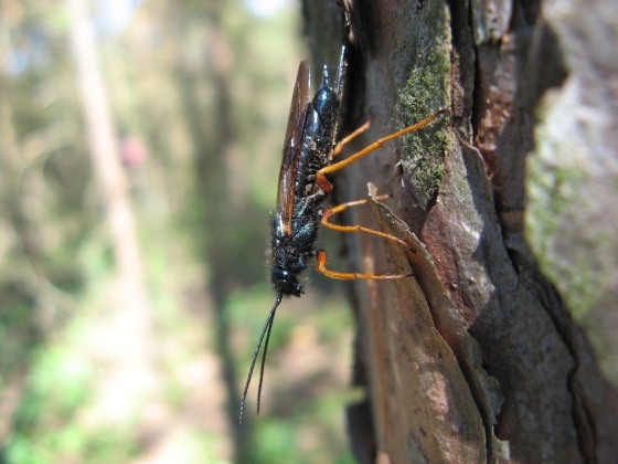
For the first issue of the current volume (145), I’ve picked the paper by Kathleen Ryan and colleagues, titled “Seasonal occurrence and spatial distribution of resinosis, a symptom of Sirex noctilio (Hymenoptera: Siricidae) injury, on boles of Pinus sylvestris (Pinaceae)“.   Sirex noctilio is a recently introduced species in Canada, and is a woodwasp that we need to pay attention to.   As Kathleen writes, “unlike our native species of woodwasps, it attacks and kills living pines” and because of this, we must strive to find effective ways to monitor the species.  One potential approach is to look for signs of resinosis, or ‘excessive’ outflow of tree sap and resins from conifers.  The goal of this work was to specifically assess “the spatial and temporal distribution of resin symptoms of attack to optimise sampling“.  The work involved Kathleen spending a LOT of time in the field, observing evidence of damage to trees, and assessing timing of resinosis relative to other damage to pine trees as related to woodwasps.  In the end, Kathleen was able to confirm that in most infested trees, the appearance of resin was a meaningful detection method.  This is a very practical paper, and very useful towards finding the best methods to detect this exotic species.

Sirex noctilio female - Photo by K. Ryan

Sirex noctilio female – Photo by K. Ryan

I asked Kathleen a few questions about this paper and the context of the work.

Q: Kathleen, what first got you interested in this area of research?

A: I became interested in studying Sirex’s interaction with other subcortical insects. Sirex was recently detected in North America at the time and we didn’t know much about it here including how, where and when to find it  – all of which were essential in planning research about insect interactions. So this study was my starting point – my “getting to know Sirex” study.

Q:  What do you hope will be the lasting impact of this paper?

A: This paper is the result of the many hours of field observations that helped me to become more familiar with Sirex. Since its really basic research, I hope that this paper might be a useful starting point for other people beginning to work with Sirex.

Q:  Where will your next line of research on this topic take you?  

A: Currently, I’m studying another invasive wood-borer, but I’d like to work with Sirex again – it’s a really interesting and unique insect biologically and ecologically. I’m especially interested in studying Sirex community ecology in its native, European, range to see how it compares to North America.

This is truly an important area of study, and I do look forward to seeing more of Kathleen’s papers in TCE.

Finally, I asked Kathleen about any amusing anecdotes about the research, and she shared this wonderful story with me:

The first day we worked together, my PhD advisor Peter de Groot, dropped me off at a forest site with instructions to only observe and collect absolutely no data. I had been in the forest for only a few moments, when a female Sirex landed right in front of me. So being an entomologist, naturally I caught her. A couple of hours later, still holding her, I met back up with Peter and sheepishly admitted that I had caught some “data”. Thinking it fantastic, from that point forward he told everyone that Sirex had picked me as her project.

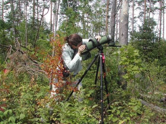
Looking for wood wasps - Photo by K. Ryan

Looking for woodwasps – Photo by K. Ryan

I believe that these kinds of stories behind the research make Entomology more accessible and real, and help us appreciate the human element of scientific research.

As a final note, the entomological community was very saddened by Peter de Groot’s death in 2010.  His legacy to Canadian Entomology is still very strong.

A special thanks to Kathleen for answering a few questions, and sharing insights into the first ‘Editor’s pick’ for The Canadian Entomologist

———-

Reference:  Ryan, K, P. de Groot, S.M. Smith and J. J. Turgeon.  Seasonal occurrence and spatial distribution of resinosis, a symptom of Sirex noctilio (Hymenoptera: Siricidae) injury on boles of Pinus sylvestris (Pinaceae). The Canadian Entomologist 145: 117-122. Link.

Female of Polistes parametricus Buck Vespidae Wasp

By Matthias Buck, Royal Alberta Museum, Edmonton

——————————————

For many of us who are working as taxonomists, describing new species has become somewhat of a routine. Sometimes it can even become a burdensome chore: I am thinking about those of us who work on hyperdiverse groups of insects in the tropics where almost every species is undescribed (case in point: one of my former lab mates recently described 170 new species of a single genus of Diptera in one paper!). However, the feeling is very different when new species unexpectedly show up in iconic groups that were thought to be well-known. Suddenly, common and familiar creatures turn into an exciting new research frontier, providing a fresh rush of adrenaline!

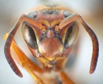
Mug shot of a female of Polistes hirsuticornis Buck. Vespidae Wasp

Mug shot of a female of Polistes hirsuticornis Buck. The hairs on the basal articles of the flagellum are longer than in related species (Photo credit D.K.B. Cheung & M. Buck).

This is what happened a few years ago when I started working on the vespids of the northeast. The family Vespidae (which includes mason wasps, paper wasps, yellowjackets and hornets) is most diverse in warmer parts of the World, as is the majority of stinging wasps. Doing a review of the northeastern Nearctic fauna therefore didn’t seem to be a very promising project for taxonomic novelty. Especially considering that the fauna of the eastern half of the continent is significantly less diverse and far better known than that of the west.

To my utmost surprise the study (published 2008 in the Canadian Journal of Arthropod Identification) not only turned up four new species of mason wasps but also two new paper wasps (Polistes). As you know, paper wasps are some of the most iconic species in the world of wasps, almost as much as their odious relatives, the yellowjackets. Further to that, they have received great attention as model organisms for the study of social behaviour and its evolution in insects. Finding not only one, but two new species in a group like this was beyond what I expected in my wildest dreams.

So how did it come to pass? As a novice to paper wasps I expected that reviewing the taxonomy of such a high-profile group would be like a walk in the park. Weren’t there scores of scientists before me who seemingly had no difficulties in identifying these sizeable and handsome insects for their behavioral studies, filling up cabinets of specimens in collections across the continent? Or so I thought! After months of fruitless staring through the microscope my nonchalant attitude gradually turned into frustration. One of the species, the common and widespread Northern Paper Wasp (Polistes fuscatus), was so variable that it blended virtually into almost every other species in the same subgenus. Previously published keys gave me a pretty clear sense of what typical specimens of each species look like, but where were the objective criteria that would allow me to identify the numerous intermediate forms? Truly, I found myself in a taxonomic quagmire!

Aedeagus of Polistes parametricus Buck. Vespidae Wasp

Aedeagus (penis) of Polistes parametricus Buck. The size, shape and position of teeth is diagnostic with regard to P. fuscatus and P. metricus, with which this species was previously confused (Photo credit D.K.B. Cheung & M. Buck).

Grasping for straws I turned to three taxonomic methods that had not been applied to Polistes before: DNA barcoding, detailed study of male genitalic features and morphometric analysis. During the previous months, I had rounded up a number of puzzling specimens which represented the spearhead of my taxonomic headaches, and submitted them for sequencing. The results came back like a thunderclap, turning my anguish into cautious excitement: the DNA barcodes of these troublemakerswere clearly different from any of the described species. With renewed energy I launched into a detailed morphological study which led to the discovery of several new diagnostic characters, confirming the distinctness of these wasps beyond a doubt. A lot of hard work had finally paid off, and I was looking at the first newly discovered species of paper wasps in eastern North America since 1836 when Amédée Louis Michel Lepeletier de Saint-Fargeau described Polistes rubiginosus!

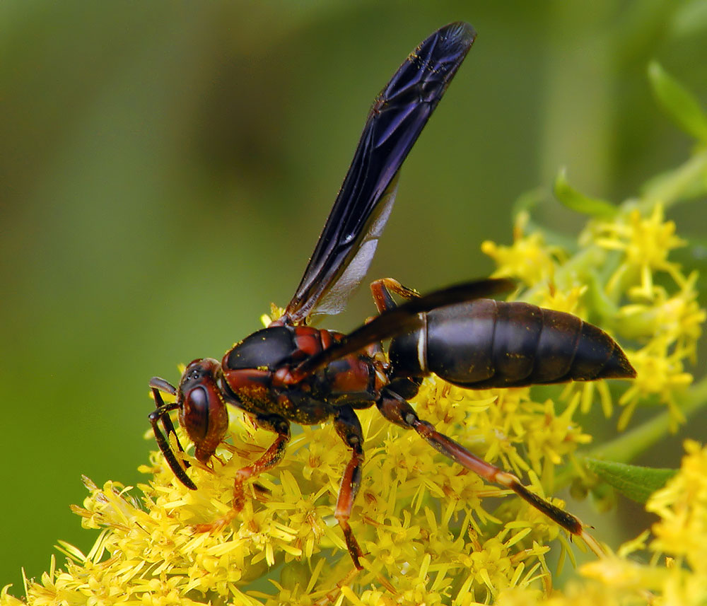
Female of Polistes parametricus Buck Vespidae Wasp

Female of Polistes parametricus Buck nectaring on goldenrod in West Virginia (Photo credit: Donna Race).

Since molecular methods, and in particular DNA barcoding, have received a lot of attention in recent years, it seems opportune to share some of my experiences working on Polistes. Unlike a few other taxa (such as spider wasps, Pompilidae), vespids sequence nicely and easily from pinned specimens, which makes them an ideal group for this kind of study. I found the sequence data extremely helpful but they certainly did not provide the cure of all taxonomic confusion. Barcoding uncovered an unexpected genetic diversity below the species level, which proved to be hard to interpret in the absence of other data. In Polistes there is no hint of a “barcoding gap”, which postulates that genetic distances between individuals of the same species are (nearly) always greater than those between conspecific individuals. In fact, some of the species were genetically so similar that they differed by a mere 2 base pairs (out of 658). Nonetheless, the combination of molecular data with fine-scale morphology resulted in a quantum leap forward for Polistes taxonomy. Just days ago, I found out that a group of researchers in Germany and Switzerland are making similar progress on European paper wasps using a nearly identical approach.

My research paper on eastern Nearctic Polistes, including formal descriptions of Polistes hirsuticornis Buck and P. parametricus Buck, was published in the journal Zootaxa on October 1st.
Matthias Buck, Tyler P. Cobb, Julie K. Stahlhut, & Robert H. Hanner (2012). Unravelling cryptic species diversity in eastern Nearctic paper wasps, Polistes (Fuscopolistes), using male genitalia, morphometrics and DNA barcoding, with descriptions of two new species (Hymenoptera: Vespidae) Zootaxa, 3502, 1-48 Other: urn:lsid:zoobank.org:pub:6126D769-A131-49DD-B07F-0386E62FF5B9

Insulin is perhaps best known as the crucial molecule whose lack leads to diabetes.  It’s a hormone that regulates carbohydrate and fat metabolism, and signals cells to increase uptake of glucose from the blood.  What most people don’t know is that insects use insulin too.

“Insulin signalling is a very conserved pathway which has been investigated extensively in humans as well as more recently in Drosophila melanogaster,” says Dr. Ana Campos, a researcher in the Biology Department at McMaster University.

And it turns out that in both insects and humans, insulin plays a much broader role in the brain than previously thought.  In a recent paper Dr. Campos and her technician Xiao Li Zhao published in the Journal of Experimental Biology, they showed that insulin signalling in the mushroom body (a critical region of the insect brain) regulates feeding behaviour in fruitfly (Drosophila melanogaster) larvae.

“Insulin has been implicated in a wide variety of biological processes. Its importance goes beyond its well-known role in the regulation of carbohydrate and fat metabolism, says Dr. Campos.  “In addition, it has been implicated in synaptic plasticity and cognitive function in humans and relevant animal models.  Recent findings indicate that abnormal insulin levels contribute to the development of neurodegenerative diseases.”

Image: Mushroom body in D. melanogaster (from Jennett et al. 2006, BMC Bioinformatics, doi:10.1186/1471-2105-7-544)

Investigating the role of insulin signalling in the mushroom body came about by a chance observation in their lab: they found a mutation in the Ran-binding protein M gene (RanBPM) that disrupted feeding behaviour in D. melanogaster larvae also inhibited insulin signalling.  Since this gene is also highly expressed in the mushroom body, it made sense to the researchers to investigate how the mushroom body influenced feeding behaviour and whether insulin signalling mediated it.

The researchers created a series of D. melanogaster strains with different parts of the known insulin-signalling pathway knocked out.  Then they measured the amount of food eaten by the different strains of mutant larvae as well as their resultant growth. By using immunohistochemical labelling, they also were able to find that reduced insulin signalling in the mushroom body on reduced the total number of neurons produced in the brains of these larvae.

Taken together, Dr. Campos and Xiao Li suggest their results mean that the mushroom body could be the brain region responsible for collecting signals about nutritional status in insects, and helps regulate feeding behaviour.  More broadly, this contributes to the knowledge about how insulin signalling impacts brain function.

Zhao, X. L. and Campos, A.R. (2012) Insulin signalling in mushroom body neurons regulates feeding behaviour in Drosophila larvae. J. Exp. Biol. http://www.ncbi.nlm.nih.gov/pubmed/22786647

Keywords: Physiology Fridays, Mushroom body, Insulin, Drosophila melanogaster, research blogging

By Brent Sinclair, University of Western Ontario
_______________________________

I’m currently on sabbatical in the Department of Zoology, University of Otago in Dunedin New Zealand.  This is the department where I did my PhD, so it is an opportunity to come back to familiar territory and re-connect with all sorts of people and places from the past.  It’s not a very insect department, but there is a lot of interesting work on ecology, parasites and freshwater biology.  A sabbatical is all about recharging scientific and creative batteries, so my main goal here is to write and read and think (and drink coffee and run and hike – but that’s for a different blog), but I felt that I also needed to justify coming all this way by actually gathering some data while I’m here.  Respirometry is the perfect answer – once set up, it’s possible to gather data on metabolic rates, breathing patterns and water loss at the expense of only a few minutes at each end of a run, leaving plenty of space for writing and drinking New Zealand’s excellent coffee in between.

What is respirometry?

Respirometry is the science (art?) of measuring the products and substrates of respiration – depending on your strategy, you can measure oxygen consumption and/or carbon dioxide production (to get a handle on metabolic rate) and water loss – among other things.  Because I work on generally small insects at generally low temperatures, we mainly measure carbon dioxide production and water loss (the instruments are much more sensitive), and can do some clever calculations to turn this into estimates of metabolic rate.

The equipment itself can look quite intimidating – and certainly like Science – with plenty of tubes and wires (when I calibrate the water channel, there’s even a bubbling flask!), but it’s not that difficult once you figure out what everything is doing, and it looks scary enough that other people generally don’t mess with it.  We pass CO2-free, dry air over an insect, and measure the CO2 and water vapour in the excurrent air – all the CO2 and water vapour must have come from the insect, so we can calculate how much it is breathing out.  The equipment we use is from a company in Las Vegas called Sable Systems International.  Sable Systems’ head honcho, John Lighton, is an insect physiologist who has published in places like Nature and PNAS, which means that when he designs the equipment, he often has insects in mind.

The respirometry system set up in a controlled-temperature room at the University of Otago. CO2-free dry air is supplied by the gas cylinder, and passes through a chamber containing the insect housed in a temperature-controlled chamber (the big grey cooler box), before going on to an infra-red gas analyser (the green box), which uses IR absorbance to measure CO2 and H2O.

What else can we learn from respirometry?

As well as a simple measure of metabolism, it is possible to use respirometry to determine the thermal sensitivity of metabolism (this is important in understanding the effects of climate change), as well as the metabolic costs of various environmental stresses, like freezing or chilling.  We can also use respirometry to study how insects breathe (there is much debate surrounding the adaptive significance of the Discontinuous Gas Exchange Cycles observed in some insects), and we can also use respirometry to figure out how much water is being lost across the cuticle of insects – even small ones like individual flies!

What am I …er… respirometing?

After 65 million years of evolution without mammals, New Zealand has an amazing array of endemism and some pretty weird insects.  My favourites are the alpine insects, which include impressive radiations of cockroaches, stick insects and weta – large Orthoptera related to the Jerusalem crickets of North America.  The mountains are fairly young (<3 million years old), so it’s possible to do all sorts of work comparing alpine species with their lowland relatives .

A group of alpine weta, Hemideina maori found under a stone at 1400 m a.s.l. on the Rock and Pillar Range, Central Otago, New Zealand. The males defend harems of 2-7 females. Female weta can weigh over 5 g, and males over 7 g, making them the heaviest insect known to survive internal ice formation. Photo by B. Sinclair.

Of course, it is the most fun to work on the big, weird insects.  So far I’ve been putting alpine weta (Hemideina maori, Orthoptera: Anostostomatidae) and New Zealand’s longest insect, the gloriously-named phasmid  Argosarchus horridus through their paces.  Male alpine weta can weigh up to 7 g, and are the largest insect species known to withstand internal ice formation.  The stick insects can easily reach 4 g, and posed some unique challenges in respirometry – with a body form so long and stick-like, it makes perfect sense to use a converted spaghetti-storage container!

A large female Argosarchus horridus (this one weighs a shade over 3 g) ready to go in her respirometry chamber. Photo by B. Sinclair.

The main questions I will be addressing will be about the evolution of thermal sensitivity and water loss in alpine insects, but the great thing about respirometry is that I never know what I’ll find along the way!

_____________________________________________________________

Brent Sinclair is an Associate Professor at the University of Western Ontario.  He is the 2012 recipient of the Entomological Society of Canada’s C. Gordon Hewitt Award.

Living in metal-contaminated lakewater is just another day’s work for phantom midge larvae. 

In the lakes surrounding Sudbury, Ontario and Rouyn-Noranda, Quebec, over 75 years of smelter operations have left their mark by contaminating soil and water with the trace metals cadmium, nickel, copper, and zinc.

This contamination led Maikel Rosabal, Landis Hare, and Peter Campbell, all from the Institut national de la Recherche scientifique in Québec, to study how aquatic animals tolerate these contaminants.  To do so, they needed a study organism that was abundant, easy to collect, and could accumulate and tolerate trace metals.  The best option turned out to be larvae of the phantom midge Chaoborus.

“The lakes in the area, and their watersheds, have been contaminated by the deposition of atmospheric aerosols and particles.  Metal concentrations in lake water tend to be higher in the lakes that are downwind from and close to the smelters, than in lakes that are upwind and distant from the smelter stacks,” says Dr. Peter Campbell. “The presence of Chaoborus in lakes with high metal concentrations implies that they are highly metal tolerant.”

The researchers chose a total of 12 lakes around Sudbury and Rouyn-Noranda with differing concentrations of trace metals, and collected water samples, using diffusion samplers that excluded particles, and midge larvae using a plankton net. After homogenizing the larvae, the researchers used a series of centrifugation, heating, and sodium hydroxide digestion steps to separate the subcellular components of the larvae.  They then measured the amount of metal in each fraction as well as the concentrations of dissolved metals in samples of lake water.  This allowed them to relate the concentrations of each trace metal in lake water to the concentrations in larvae.

They found that the majority of each metal accumulated in the cytosolic heat-stable protein fraction that they isolated from the larvae—a fraction that contains large amounts of metal-binding proteins. And while other fractions also contained small amounts of metals, it was in the heat-stable protein fraction that metal concentrations responded most obviously to the increasing metal concentrations in lake water. This suggests that the Chaoborus larvae were able to bind and detoxify increasingly large amounts of these potentially toxic metals.

“This suggests an important role for these metallothionein-like proteins in the detoxification of metals,” says Dr. Campbell.  “Presumably this contributes to the presence of this insect in highly metal-contaminated lakes.”

While laboratory studies usually focus on the effects of exposure to a single trace metal (usually dissolved in the water), animals in this study were exposed in the field to many trace metals both in the water and in their planktonic food. The researchers suggest that Chaoborus larvae would be effective “sentinels” for estimating trace-metal exposure to lake plankton, which is a key component of ecological risk assessments.

“Rough estimates of trace metal exposure are often obtained by measuring total metal concentrations in the water or the sediment.  Such values usually overestimate metal exposure because much of the metal present is not available for uptake by organisms because they are bound to substances such as organic matter or iron oxides,” explain the researchers. “For these reasons, measurements of trace metals in organisms are increasingly used to estimate exposure in risk assessments.”

Rosabal, M., Hare, L. & Campbell, P.G.C. (2012). Subcellular metal partitioning in larvae of the insect Chaoborus collected along an environmental metal exposure gradient (Cd, Cu, Ni and Zn), Aquatic Toxicology, 120-121 78. DOI: 10.1016/j.aquatox.2012.05.001

Pubmed: http://www.ncbi.nlm.nih.gov/pubmed/22647479

Chaoborus larvae

Photo: Maikel Rosabal

Chris Buddle, Editor-in-Chief, The Canadian Entomologist
________________________________

I’ve been involved with the Entomological Society of Canada for a long time.  It’s a wonderful community of Canadian entomologists sharing an interest and enthusiasm for arthropods. The ESC’s activities are mostly centered around  its annual conference, its range of publications, and it offers a suite of awards and scholarships.  The society’s website also hosts career opportunities, photo contests, and a range of other rich and varied entomological content. The latest, big news for the society is that on 1 June, the ESC officially launched its own blog.  This blog was the brainchild of a few members of the society.

So…why does a scientific society need a blog?  What’s the benefit to members of the society, to the society itself, and what’s the benefit for the broader entomological community?  Here are some thoughts about this:

1) Visibility:  it’s a tough time for scientific societies – funding is tight, and for a lot of people, the value of memberships to societies may seem less important than it once was.  Therefore, increased visibility though an on-line presence is important. A static website is essential, but a blog has a fluidity and dynamic presence that is hard to match with a website.  An active blog with well-written and interesting content will do a lot to increase a society’s visibility.  The visibility from an active blog is also global in its reach.

2) Opportunities to contribute:  the ESC blog will have dozens of contributors – means anybody with an interest in entomology (regardless of their profession and educational background) has an opportunity to write something for a broader audience.  Blog posts are often easier to write, they are shorter than research papers, and the content need not be vetted through a peer-review process.  It’s a forum for creative ideas, stories, photographs, and fun facts about insects.  The blog already has a couple of nice examples to illustrate this point.  For example, Chris Cloutier, a naturalist at the Morgan Arboretum on the Island of Montreal, just wrote a lovely post about the Hackberry Emperor.  Chris is an example of a different kind of entomologist – he’s not a research scientist, nor is his primary profession Entomology.  However, he does outreach, has a wealth of expertise and  talent, and he has a lot to offer the entomological community.  These kind of opportunities create an environment of inclusion for a society – members have a voice and can share their ideas and expertise.  Non-members can also contribute and recognize that there is a strong community associated with the ESC (…and perhaps some of the non-members will see the value of the society and join).

3) Economics: more than ever before, scientific societies are struggling to maintain members, and balance their books.  A blog is a cheap and effective way to promote their science to the world and the cost can be as little as a domain name.  I can think of no other method by which a society can promote itself at this cost point.  You could even argue that the time for static websites may be coming to a close since they are costly to host, require people with specific technical skills, and require a lot of back-end support.  The good blog sites can be administered by people with relatively few of these skills (I’m proof of that!!).

4) Marketing and branding:  a high quality blog helps a society get its brand to a broad audience, and helps to market the society to the world.   The ESC has a long and wonderful history, but its main audience over the years has mostly been academics, research scientists, and students of entomology.   The ESC brand has excellence and quality behind it and that kind of brand should be shared, expanded, and through this process, the society will hopefully gain positive exposure and more members.

5) Communication: At the end of the day, knowledge is something to be shared.  Scientific communication is a fast-changing field and one that is making all of us reconsider how we talk and write about our interests.   I think we all have a responsibility to do outreach.  There is so much mis-information out on the Internet, and people with specialized and well-honed skills must be heard and must have a means to share accurate information in a clear and effective manner – e.g., a society blog. I also think many entomologist are perfectly positioned to do effective outreach (I’ve written about this before).  Part of the ESC’s mandate is dissemination of knowledge about insects and social media is a key piece of any communication strategy.

What do you think?  Can you think of other reasons why scientific societies need to embrace social media?  Please share your ideas!

I will finish with a stronger statement:  scientific societies are perfectly positioned to have the BEST blogs on the Internet.  A scientific society is a community, a community with history, and a community built on high level of expertise.  A scientific society also provides a structure and framework for bringing together high quality knowledge about a particular topic.  A blog can be amazingly strong with this kind of support.  A society is also about people and these people work tirelessly behind the scenes to facilitate the dissemination of high quality content.   These people, structured in committees, and with oversight from an executive committee, can provide tangible support that will help to keep a blog from becoming unidimensional.  The ESC’s blog administrators (Crystal and Morgan) know how to keep the content of high quality, and know how to put all the pieces together – and they know they can do this because they have an entire community behind them.  The society is committed to supporting the blog and for that reason, there is reason to be optimistic about its long-term success.

___________________________

Originally posted at: http://arthropodecology.com/2012/06/13/why-a-scientific-society-needs-a-blog/

Dufourea bee on flower

pcyu_logo

By Sheila Dumesh, entomology research assistant at York University.

——————————–

My interest in bees was ignited in 2007, when I took a biodiversity course in my last year as an undergraduate student at York University in Toronto.  The course instructor was the well-known melittologist, Laurence Packer, and, although I had not met him before, I had heard many good things.  Laurence’s affection for bees was inspiring, not only to me, but to others in the past and many more to come.  He was so fascinated by these cute and fuzzy insects (at the time, I did not see myself describing them as such).  Even though he had been studying bees for decades, the look of excitement on his face never faded when collected and examined them.  Back then, my knowledge of bees was very limited.  I was unaware of their diversity, importance, and great beauty!

I began with an Honours thesis under Laurence’s supervision in the “bee lab” at York University.  I was keen on taxonomy and began a systematic study on a Central American bee genus, Mexalictus.  For my Master’s thesis, I chose to continue that work and complete a revision of Mexalictus, which included descriptions for 20 new species, an illustrated key, and a phylogenetic analysis.  I conducted my field work in Costa Rica, Guatemala, and Mexico, where I sampled in high elevation cloud forests (the known habitat of Mexalictus).  As these species are quite rare, I did not always have the pleasure of finding them; although this was somewhat upsetting, I was amazed by the bee (and general insect) diversity in that part of the world.  I was aware of it, but being out in the field in those countries was a truly amazing experience.  Just the change in habitat and species make-up along a small sector of the elevation gradient was incredible to witness!

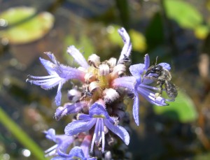
Dufourea bee on flower

Dufourea sp. – Photo by Sheila Dumesh

Throughout my time as a Master’s student, I studied other groups of bees and collaborated with others in our lab.  One such project is the revision of the Canadian species in the genus Dufourea (Apoidea: Halictidae), which I undertook with Cory Sheffield and recently published in the Canadian Journal of Arthropod Identification.  There are eight species in Canada, but some were described from only one sex, the descriptions were written by several authors in different publications, and a key to identify these species was previously unavailable!  These bees are also floral specialists, meaning they visit specific flowers (usually a genus or family).  Cory and I set out to revise this group and provide all of this information in one paper.  The identification key is user-friendly and illustrates the characters mentioned in the key couplets to aid the user.  We also constructed species pages, which include full descriptions, important features, distribution maps, and images of each species.

We are striving towards creating many more illustrated (and web-based) keys to facilitate bee identification.  I am very excited to have this work freely available and hope that it is found useful by others in the community!

———–

Dumesh, S. & Sheffield, C.S. (2012). Bees of the Genus Dufourea Lepeletier (Hymenoptera: Halictidae: Rophitinae) of Canada, Canadian Journal of Arthropod Identification, 20 DOI: 10.3752/cjai.2012.20

Honey bee flying with pollen - Photo by Alex Wild

Honey bee flying with pollen – Photo by Alex Wild, used with permission

Honeybee colonies are famous for their orderly divisions of labour.  As worker bees grow up, they transition from housekeepers (cleaning the colony) to nurse bees (feeding young bees), to finally switching to foragers who go out and collect nectar and pollen for the rest of the colony.   To maintain a healthy colony, bees need to decide how many foragers and how many nurse bees are needed, and control of these numbers is accomplished by pheromone levels within the colony.

In honeybee colonies, there are pheromones like the alarm pheromone that cause immediate behavioural responses (called releaser pheromones) and others that trigger physiological changes like hormones do (called primer pheromone).  From previous work, it seemed that ethyl oleate functions as a primer pheromone, produced by foragers, that delays the maturation of nurse bees into foragers.

“Ethyl oleate does not elicit any noticeable behavourial responses in recipient workers,” says Dr. Erika Plettner, who supervised a recent study on the synthesis of ethyl oleate at Simon Fraser University in British Columbia.  “Yet it has a profound physiological effect”.

To understand how this chemical is produced in the individual bee and then distributed in the colony, Carlos Castillo and colleagues from Simon Fraser University in British Columbia and the Laboratoire Biologie et protection de L’Abeillie in France looked at several ways to identify the source and synthesis of ethyl oleate.  This chemical can be produced by a reaction between oleic acid (a common fatty acid in insects) and ethanol.  While you might not think of honeybees as heavy drinkers, it turns out that yeasts in flower nectar ferment the sugars present into ethanol, and so the forager bees have much higher exposure to ethanol than nurse bees.

To figure out if ethanol and oleic acid can be made into ethyl oleate by honeybees, the researchers incubated different honeybee body parts from forager and nurse bees with these precursors.  They found highest production of ethyl oleate in the head tissues, and that both nurses and foragers could produce ethyl oleate when given ethanol.  In addition, in whole bees, they found that the ethyl oleate migrated from the gut to the exoskeleton of the bees where it would exude into the colony.

Taken together, these results suggest that making ethyl oleate, while it is useful for colony control, might also be a way to deal with the occupational hazard of consuming toxic ethanol.  “Foragers have much higher occupational exposure to ethanol than nurses do,” says Dr. Plettner.  “This is why they make ethyl oleate in nature”.

Ethyl oleate molecule

Ethyl oleate

To track down where exactly the ethyl oleate was synthesized, they coupled oleic acid to a chemical that would produce fluorescence when the oleic acid was combined with ethanol to produce ethyl oleate.  Under the microscope, areas that fluoresced showed where ethyl oleate was being made.  They found that ethyl oleate was made in the esophagus, honey crop and stomach.

The authors were also able to identify the genes responsible for the synthesis of ethyl oleate in the honeybee and the resulting enzymes that catalyze the reaction between oleic acid and ethanol.  These enzymes are then secreted into the gut fluid, where they produce ethyl oleate, which is then transported to the cuticle.

The biosynthesis of ethyl oleate then can be thought of a way of providing updates to the colony about the availability of flower nectar in nature.  “EO might be some kind of ‘resource meter’ that tells the nurses in the colony how many nectar and pollen resources are coming in,” says Dr. Plettner.  “If lots of food is coming in, then it makes sense to inhibit nurse to forager transition, as the nurses would be more needed in the brood chamber than as foragers.  Conversely, if few resources and/or foragers are coming in, then it makes sense to speed up development of nurses so that they can forage and fill the need.”

Castillo, C., Chen, H., Graves, C., Maisonnasse, A., Le Conte, Y. & Plettner, E. (2012). Biosynthesis of ethyl oleate, a primer pheromone, in the honey bee (Apis mellifera L.), Insect Biochemistry and Molecular Biology, 42 (6) 416. DOI: 10.1016/j.ibmb.2012.02.002

Corresponding author: Erika Plettner (plettner@sfu.ca)

Further reading:

Castillo, C., Maisonnasse, A., Conte, Y.L. & Plettner, E. (2012). Seasonal variation in the titers and biosynthesis of the primer pheromone ethyl oleate in honey bees, Journal of Insect Physiology, 58 (8) 1121. DOI: 10.1016/j.jinsphys.2012.05.010

Pollenia griseotomentosa Calliphoridae Cluster fly
Pollenia rudis Face

Pollenia rudis

By Adam Jewiss-Gaines,  a research assistant at Brock University.

———————

When people ask me what the heck a calliphorid is (often after I have mentioned the family name and am being gawked at as if I’m crazy), I usually remark “You know those shiny flies you often see flying around in the spring and summer?”  This isn’t technically 100% accurate since the genus Pollenia, one of the most commonly encountered genera of the family, is in fact non-reflective and grey.  Upon closer inspection, a keen eye can also observe varying amounts of wrinkled, yellow hairs on the thorax.  These two qualities distinguish Pollenia from other blow flies throughout North America.  Despite being a little dull when compared to their more eye-catching iridescent relatives, Pollenia are ecologically important insects as they aid in plant pollination and the processing of various biomaterials.

Pollenia often become particularly active during the spring and summer months once the temperature warms up, although they can occasionally be spotted indoors in the wintertime on a warmer day.  With a sudden onslaught of large, grey insects flying around when the snow begins to melt, it comes as no surprise that people tend to get irritated with them and consider them pests.  Oftentimes they are mistaken as houseflies (Family Muscidae) causing Pollenia species to be labeled as potential food contaminators, but this is not the case.  These insects are also particularly well-known for their clustering behaviour on walls, earning them their common name: cluster flies.

Even though Pollenia are extremely common, their general biology is largely unknown with a few exceptional details. It is known that larval Pollenia are parasites on various other organisms, such as maggots and worms. For example, Rognes (1991) noted that Pollenia pediculata, one of the most common species found throughout the continent, is a parasite of the earthworm species Eisenia rosea. Aside from this little tidbit however, specific information regarding the life cycles of Pollenia species is relatively scarce and further studies in this particular field would greatly improve our knowledge of the genus.

Pollenia griseotomentosa Calliphoridae Cluster fly

Pollenia griseotomentosa

Until very recently it has been thought that all Pollenia found in North America were the same species (Pollenia rudis), but after examining various collections throughout the world, Knut Rognes found that six members of the genus occur throughout the region.  Terry Whitworth adapted much of Rognes’ work shortly thereafter into a nice, clean, simple identification key for North America. With accurate images and photography, however, characters could be even easier to distinguish and observe when one is able to compare a photograph to the creature they have under their microscope.

Therefore, to further expand on Terry’s key and clarify important visual characters, I collaborated with him and Dr. Steve Marshall to create a fully-illustrated digital key for distinguishing the six North American Pollenia species from one another.  Now published in the Canadian Journal of Arthropod Identification, Cluster Flies of North America couples high-resolution images of important traits with a clean and simple interface to create a handy tool to be used by entomologists and non-entomologists alike. If you are relying on this key for identification, it is recommended to use physical specimens of Pollenia rather than images or photos, since even the best of hand-photographs have difficulty capturing key features. In addition, distribution maps are provided for each species, constructed from locality data of specimens from the University of Guelph Insect Collection and Terry Whitworth’s personal collection of Pollenia.

Creating this key has been a great opportunity, and I hope the entomological community is able to make good use of it. My sincere thanks go out to Steve Marshall, Terry Whitworth, the editors, and my labmates and friends for all of their support.

——————————

Jewiss-Gaines, A., Marshall, S.A. & Whitworth, T.L. (2012). Cluster flies (Calliphoridae: Polleniinae: Pollenia) of North America, Canadian Journal of Arthropod Identification, 19 DOI: 10.3752/cjai.2012.19

Rognes, K. 1991. Blowflies (Diptera, Calliphoridae) of Fennoscandia and Denmark. Fauna Entomologica Scandinavica Vol. 24.

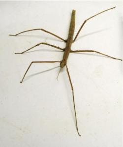
Stick Insect Baculum extradentatum

Physiology Friday is a monthly column by UWO PhD candidate Katie Marshall and will feature new Canadian research on insect physiology.

——————

Nitric oxide (NO) is usually overshadowed in fame by its more famous cousin laughing gas, but it’s difficult to think of many simple molecules that have such a variety of important biological functions.  While NO only lasts a few seconds in the free gaseous state in the blood, it has been implicated in processes that involve everything from immune function to neurotransmission.  One important role for NO is in the cardiac system, where it functions as a vasodilator and in vertebrates it slows heart rate, while in insects it has the opposite effect.

Stick Insect Baculum extradentatum

Baculum extradentatum photo by Sara da Silva

Most of the research about the physiological functions of NO has focused on vertebrates, but recent work published in the journal of Cellular Signalling by graduate student Sara da Silva and her postdoctoral fellow mentor Rosa da Silva in the lab of Angela Lange (University of Toronto Mississauga), has shown that, unlike other insects, the Vietnamese stick insect Baculum extradentatum can respond to NO like a vertebrate.

“Our initial research interests in cardiac physiology were influenced by earlier work indicating that stick insect hearts are innervated and can be modulated by endogenous chemicals [like NO],” says study director and University of Toronto Biology professor Angela Lange.  “It is for this reason that we chose this understudied organism, which contains a simplified cardiovascular system that can be considered a model for work on other cardiac systems.”

The researchers first attempted to find the natural source of NO in the stick insect by removing hemolymph (blood) samples and staining for the presence of an enzyme that produces NO.  Then they examined the effects of NO on heart rate by dissecting the dorsal vessel out and maintaining it in a Petri dish with physiological saline.  They could measure heart rate through the placement of electrodes on either side of the dissected heart, and monitor the effects of various chemicals on the cardiac activity of the stick insect.   They also could examine whether heart rate was mediated by the central nervous system by leaving the nervous system attached or not.

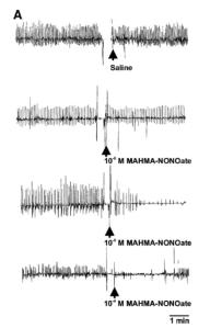
insect heart rate

The effects of nitric oxide on the heart rate of B. extradentatum. Figure 3 from da Silva et al. 2012

They found that the hemocytes (blood cells) of the stick insect were producing an enzyme that was similar to the enzyme other animals use to produce NO.  In addition, the more of a chemical called MAHMA-NONOate (which produces NO) they added, the slower the stick insect hearts beat.  This surprising effect was completely opposite to what had been found in other insects and was more like the response of the vertebrate heart.

“Insects have evolved different strategies depending upon life history, and have co-opted different messenger systems for this success,” says study author da Silva. “We need to understand the full ecology of all species to finally appreciate the factors involved.”

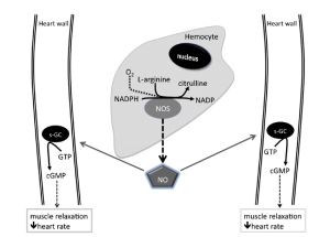
Using the same setup, they also tested other components of a system of compounds that they thought might be involved in the pathway that produces NO that leads to decreased heart rate in B. extradentatum.  They believe that NO is produced in the hemocytes, travels to the wall of the heart, and then leads to the production of a messenger molecule that decreases heart rate.

Schematic diagram of the proposed regulation of cardiac activity in B. extradentatum by the gaseous signaling molecule, nitric oxide (NO)

Schematic diagram of the proposed regulation of cardiac activity in B. extradentatum by the gaseous signaling molecule, nitric oxide (NO). Figure 7 from da Silva et al. 2012.

“This study further emphasizes the evolutionary links between the physiological processes of vertebrate and invertebrate systems,” says da Silva. “Our findings suggest that signaling molecules (such as NO) common to both types of organisms can have similar effects on cardiac activity.  These novel findings demonstrate that the study of vertebrate systems can be complemented with studies in model invertebrate organisms.”

—————————-
da Silva, R., da Silva, S.R. & Lange, A.B. (2012). The regulation of cardiac activity by nitric oxide (NO) in the Vietnamese stick insect, Baculum extradentatum, Cellular Signalling, 24 (6) 1350. DOI: 10.1016/j.cellsig.2012.01.010