Female of Polistes parametricus Buck Vespidae Wasp

By Matthias Buck, Royal Alberta Museum, Edmonton

——————————————

For many of us who are working as taxonomists, describing new species has become somewhat of a routine. Sometimes it can even become a burdensome chore: I am thinking about those of us who work on hyperdiverse groups of insects in the tropics where almost every species is undescribed (case in point: one of my former lab mates recently described 170 new species of a single genus of Diptera in one paper!). However, the feeling is very different when new species unexpectedly show up in iconic groups that were thought to be well-known. Suddenly, common and familiar creatures turn into an exciting new research frontier, providing a fresh rush of adrenaline!
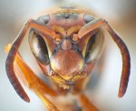
Mug shot of a female of Polistes hirsuticornis Buck. Vespidae Wasp

Mug shot of a female of Polistes hirsuticornis Buck. The hairs on the basal articles of the flagellum are longer than in related species (Photo credit D.K.B. Cheung & M. Buck).

This is what happened a few years ago when I started working on the vespids of the northeast. The family Vespidae (which includes mason wasps, paper wasps, yellowjackets and hornets) is most diverse in warmer parts of the World, as is the majority of stinging wasps. Doing a review of the northeastern Nearctic fauna therefore didn’t seem to be a very promising project for taxonomic novelty. Especially considering that the fauna of the eastern half of the continent is significantly less diverse and far better known than that of the west.

To my utmost surprise the study (published 2008 in the Canadian Journal of Arthropod Identification) not only turned up four new species of mason wasps but also two new paper wasps (Polistes). As you know, paper wasps are some of the most iconic species in the world of wasps, almost as much as their odious relatives, the yellowjackets. Further to that, they have received great attention as model organisms for the study of social behaviour and its evolution in insects. Finding not only one, but two new species in a group like this was beyond what I expected in my wildest dreams.

So how did it come to pass? As a novice to paper wasps I expected that reviewing the taxonomy of such a high-profile group would be like a walk in the park. Weren’t there scores of scientists before me who seemingly had no difficulties in identifying these sizeable and handsome insects for their behavioral studies, filling up cabinets of specimens in collections across the continent? Or so I thought! After months of fruitless staring through the microscope my nonchalant attitude gradually turned into frustration. One of the species, the common and widespread Northern Paper Wasp (Polistes fuscatus), was so variable that it blended virtually into almost every other species in the same subgenus. Previously published keys gave me a pretty clear sense of what typical specimens of each species look like, but where were the objective criteria that would allow me to identify the numerous intermediate forms? Truly, I found myself in a taxonomic quagmire!

Aedeagus of Polistes parametricus Buck. Vespidae Wasp

Aedeagus (penis) of Polistes parametricus Buck. The size, shape and position of teeth is diagnostic with regard to P. fuscatus and P. metricus, with which this species was previously confused (Photo credit D.K.B. Cheung & M. Buck).

Grasping for straws I turned to three taxonomic methods that had not been applied to Polistes before: DNA barcoding, detailed study of male genitalic features and morphometric analysis. During the previous months, I had rounded up a number of puzzling specimens which represented the spearhead of my taxonomic headaches, and submitted them for sequencing. The results came back like a thunderclap, turning my anguish into cautious excitement: the DNA barcodes of these troublemakerswere clearly different from any of the described species. With renewed energy I launched into a detailed morphological study which led to the discovery of several new diagnostic characters, confirming the distinctness of these wasps beyond a doubt. A lot of hard work had finally paid off, and I was looking at the first newly discovered species of paper wasps in eastern North America since 1836 when Amédée Louis Michel Lepeletier de Saint-Fargeau described Polistes rubiginosus!
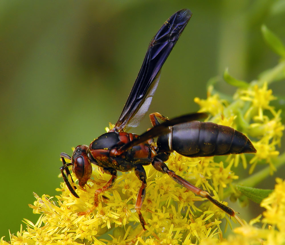
Female of Polistes parametricus Buck Vespidae Wasp

Female of Polistes parametricus Buck nectaring on goldenrod in West Virginia (Photo credit: Donna Race).

Since molecular methods, and in particular DNA barcoding, have received a lot of attention in recent years, it seems opportune to share some of my experiences working on Polistes. Unlike a few other taxa (such as spider wasps, Pompilidae), vespids sequence nicely and easily from pinned specimens, which makes them an ideal group for this kind of study. I found the sequence data extremely helpful but they certainly did not provide the cure of all taxonomic confusion. Barcoding uncovered an unexpected genetic diversity below the species level, which proved to be hard to interpret in the absence of other data. In Polistes there is no hint of a “barcoding gap”, which postulates that genetic distances between individuals of the same species are (nearly) always greater than those between conspecific individuals. In fact, some of the species were genetically so similar that they differed by a mere 2 base pairs (out of 658). Nonetheless, the combination of molecular data with fine-scale morphology resulted in a quantum leap forward for Polistes taxonomy. Just days ago, I found out that a group of researchers in Germany and Switzerland are making similar progress on European paper wasps using a nearly identical approach.

My research paper on eastern Nearctic Polistes, including formal descriptions of Polistes hirsuticornis Buck and P. parametricus Buck, was published in the journal Zootaxa on October 1st.
Matthias Buck, Tyler P. Cobb, Julie K. Stahlhut, & Robert H. Hanner (2012). Unravelling cryptic species diversity in eastern Nearctic paper wasps, Polistes (Fuscopolistes), using male genitalia, morphometrics and DNA barcoding, with descriptions of two new species (Hymenoptera: Vespidae) Zootaxa, 3502, 1-48 Other: urn:lsid:zoobank.org:pub:6126D769-A131-49DD-B07F-0386E62FF5B9

Vivez une expérience audio visuelle hors du commun en compagnie du conférencier Étienne Normandin. Musiques, sons et vidéos de la BBC et de « Bugs of the underworld » sont au menu dans le but d’émerveiller vos sens à la beauté et à la fantastique entomofaune aquatique.

Les insectes aquatiques ont été les premiers insectes à apparaître sur la Terre, mais aussi les premiers à utiliser la voie des airs. Dans cette conférence dédiée à un public de tous âges, vous en apprendrez plus sur les particularités des insectes aquatiques ainsi que leurs comportements. Ces insectes peuvent être de fameux architectes, des pêcheurs habiles et d’excellents chasseurs. Ils sont aussi très importants pour l’écologie des plans d’eau et sont de bons outils pour les biologistes.

2 novembre 2012, 19h00

Jardin Botanique, IRBV, Local 354

link: www.aeaq.ca
page facebook: Association des entomologistes amateurs du québec
groupe facebook: Association des entomologistes amateurs du québec

The Entomological Society of Canada is looking for volunteers for the upcoming JAM, November 3-7!

Volunteering looks great on your CV, is an excellent way to meet new people, and is fun! The Student Affairs Committee worked hard to keep student registration rates low, so we need a very strong showing of student volunteers to help make this meeting a success!

Sign up at http://www.doodle.com/i8znn4z75mtharfw by checking off times you are available. The full program is up now so you can confirm when you are presenting: check it out here!

A friendly reminder from  Adrian Thysse about the JAM 2012 Photo Competition!
_____________________________
The deadline for submission to the 2012 JAM photo competition is October 30! All  JAM 2012 participants are eligible to enter, so why not submit your best shots now?

The theme for the competition will be Canadian Arthropods, and there are four categories:

  1. Dead – pinned or preserved specimens
  2. Alive – in the natural habitat
  3. Dead or Alive – predators with prey
  4. Alive with mites – insect mite symbiosis (Sponsored by International Journal of Acarology editor, Dave Walter)

$150 will be awarded to the winner for each category and the “Alive with mites” winner may be offered the opportunity to be a cover illustration for the International Journal of Acarology.

So far the judges include John Acorn, David Walter and myself, and we are looking forward to a wealth of submissions from all the many entomologists, amateur or professional, that will be attending JAM 2012.

Go check out the rules and submit today!

The results of the Eighth Annual ESC Photo Contest have been announced!  Judges Kirk Hillier, Kenna MacKenzie, and Rick West faced a difficult task, selecting the winners from among 67 high-quality entries.

The top seven selection will be printed on the cover of all issues of Volume 145 (2013) of The Canadian Entomologist.  The photos were chosen primarily for their composition and quality, but judges also tried to spread the winning entries across insect orders, and to have no more than one winning photo per photographer. The final results are:

First Place: Bob Lalonde, “Halictid on fireweed”. A female Agapostemon sp. (Halictidae), foraging on fireweed in June on the UBC Okanagan (Kelowna) campus.

Second Place: Ward Strong, “Stinkbug eggs”. Stinkbug eggs found on the foliage of lodgepole pine, Tappen BC.

Third Place: Julian Dupuis, “Papilio larva on Artemesia”. Larva of Papilio machaon dodi (Lepidoptera: Papilionidae), on Artemesia dracunculus, near Drumheller, AB

Fourth Place: Crystal Ernst, “Stratiomys badia”. An impressive bee mimic, Stratiomys badia (Stratiomyidae) rests in a garden at dusk, in Chesterville, Ontario.

Fifth Place: John McLean, “Honeybee Drone pupae”. Late stage pupae of the honey bee Apis mellifera L. dissected as part of a search for breeding varroa mite (none found). Taken from a hive in the Gisborne area on the East Coast of the North Island of New Zealand, March 2012.

Sixth Place: Tim Haye, “Pachycoris klugii nymphs”. Nymphs of Pachycoris klugii on Jatropha cucras tree (Tehuacan, Chiapas, Mexico).

Seventh Place: Christa Rigney, “Dakota Skipper on Yarrow”. A gravid female of the Threatened Dakota skipper, Hesperia dacotae (Skinner) (Hesperiidae) perched on Yarrow, Achillea millefolium (L.) (Asteracea) in a tallgrass prairie northeast of Deleau, Manitoba

A slideshow of all of the beautiful photographs entered in this year’s Competition is now displayed on the ESC Website, here. Congratulations to the winners!!!

If you missed this year’s competition, don’t fret! There is still time to submit your own images to another ESC-sponsored photo contest! ESC (or other regional society) members attending this year’s Joint Annual Meeting in Alberta have until October 30th to get their best shots of the year in to the judges of the 2012 JAM Photo Contest.

Thank you one and all for your participation, and keep those shutters clicking!

Hi, my name is Holly Caravan and I am a PhD student in Dr. Tom Chapman’s social insect lab at Memorial University of Newfoundland. Currently my work is focused on galling aphids and their potential for antimicrobial activity within the gall. This past summer I visited Dr. Patrick Abbot’s lab at Vanderbilt University (Nashville, TN) where I was able to access three species of galling aphids. But, to address the ultimate goal of my research, I want to include the species Pemphigus spyrothecae which produces spiral galls on Lombardy poplar, Populus nigra. This species has a soldier caste which is morphologically specialized, different from the other three species I have already researched. I am looking for any information on locations of this aphid species in Canada; Newfoundland would be ideal, but my hopes are not high! Attached are links with pictures of the host tree and the spiral galls produced by the aphids. Any information would be greatly appreciated! I can be contacted at holly.caravan@gmail.com or hcaravan@mun.ca!

http://www.naturespot.org.uk/species/pemphigus-spyrothecae

http://www.parkwoodpines.com.au/html/lombardy_Poplar.html

Jacob Coates is an MSc student in the Chapman Entomology Lab at Memorial University of Newfoundland and Labrador.

—————————————

Cockroach – Photo by Jacob Coates

If you’ve never thought of visiting Australia, you’re making a terrible mistake. I just recently returned from a 6 month stint in Sydney based out of a Lab in Macquarie University. I carried out lab and field work on several species of gall-inducing thrips. I owe this great trip to the Australian Endeavour’s Awards, An Australian government run program which takes applications from students all over the world and to those lucky enough to be accepted, ships you to an Australian University with a wage, living allowance and travel cash. On top of getting some serious work done I enjoyed snorkeling around the many beaches, hiking in the Blue Mountains, and took part in the City 2 Surf road race where over 80,000 individuals take to the streets of Sydney to run the largest road race in the world.

Southern Queensland Red Road – Photo by Jacob Coates

In early June I completed a field trip into Southern Queensland to collect insect samples. Tenting through the outback presented some difficulties like torrential downpours, cold nights, and very sloppy road conditions (Nearly sinking a 4×4 in a flooded dirt road). Despite the problems, after nearly 2500 kms and 10 days of driving I returned to Sydney with thrips samples in hand and a very dirty truck to clean. Amazing wildlife, epic landscapes and great people await everyone in the outback, without a doubt the best trip of my life.

Jacob Coates

For those interested about the Endeavour’s award go to http://www.deewr.gov.au/International/EndeavourAwards/Pages/Home.aspx It’s well worth your time.

Another close vote for Photo 3 captions, but ultimately Brian Cutting took home top spot and 5 points, while Greg & Colin tied for 2nd and earned 3 points each. We’ll tally up the scores next time at the half-way point, and unveil the prizes, but there’s still plenty of time for you to join in!

With only 3 captions for Photo #4, everyone’s guaranteed points, but it’s up to you to decide who earns the top honours:

[polldaddy poll=6591094]

This week’s photo is a little different, but we’re sure you can dazzle us with your wit! (Rules)

Photo by Morgan Jackson

Culex pipiens photo by Kate Bassett

Today’s post is by Kate Bassett of Memorial University. If you’d like more information about her work, she encourages you to contact her.

——————————-

Hi,

I’m a graduate student at Memorial University (MUN, St. John’s, Newfoundland and Labrador), nearing the end of my masters…hopefully :). My research project is focused on a wildlife issue. Snowshoe hare, Newfoundland’s only Lagomorph, suffer from infection by California serogroup viruses (snowshoe hare virus and Jamestown Canyon virus). Helped by the province’s Chief Veterinarian Officer Dr. Hugh Whitney, I sampled the blood and tested for infection in wild hares and laboratory rabbits used as sentinels.  This work was based in part in the laboratory led by microbiologist Dr. Andrew Lang at MUN, as well as working with the team at the National Microbiology Lab headed by Dr. Michael Drebot in Winnipeg. But, my project also included studying mosquitoes that are thought to transmit these viruses. That part of my project was based in the social insect lab at MUN headed by Dr. Tom Chapman.

I spent two summers catching mosquitoes. Consequently, I can’t miss them. I seem to have permanently altered my hearing and vision such that a mosquito in flight always grabs my attention. Last May while putting in a load of laundry, a specimen alighted on the washer. I dropped everything and ran upstairs for my aspirator, and made it back to collect this girl to identify at work. I froze her and didn’t get around to id’ing until later in the summer, and I was shocked to see that it may be Culex pipiens. This mosquito gains attention on the East Coast of North America because it can transmit West Nile Virus, and when I made this determination the worst West Nile viral outbreak in N.A. was underway and centered in Texas. I was uncertain of my morphological identification, so I added a leg or two of this specimen to my DNA barcoding work, and I waited for the outcome. When the sequence confirmed by identification, I put out a press release, which had me immediately doing live interviews on TV and Radio. I didn’t have a lot of time to think about it, I just went from interview to interview. It was a good experience; I do recommend it. I should add that we don’t have confirmation of West Nile Virus in Newfoundland, but we don’t know what lies ahead. Drs. Lang (aslang@mun.ca), Chapman (tomc@mun.ca) and Whitney (hughwhitney@gov.nl.ca) are looking for students to pick up where I am leaving off.

Culex pipiens photo by Kate Bassett

Here’s a picture of Cx. pipiens I took using a digital camera mounted on a dissecting scope. I used the program Helicon for producing a wide focal plane. It’s not the one that I got in May and fingerprinted, but another one that I got last weekend (September, 2012), also in my house!